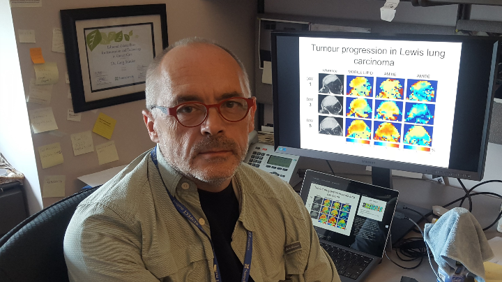
TFRI-funded researchers based in Toronto have found that a specific type of magnetic resonance imaging (MRI) tool may be able to measure how breast cancer patients respond to chemotherapy within a day of treatment.
Published in Oncotarget (July 2018), the finding reveals that chemical exchange saturation transfer MRI (CEST MRI) can detect cancer cell death in mice within 24 hours of receiving chemotherapy, much sooner than current methods, which measure changes in tumour size and may take up to six months to reveal if patients effectively respond to treatment.
There are no clinically used methods now for detecting early tumour responses to therapy. The team sought to explore new tools because current techniques have significant limitations pertaining to reliability, sensitivity, specificity and, in some cases, cost more to use. This tool “does not require injections of exogenous media and could be easily integrated into existing clinical MRI protocols,” state the study authors.
“This finding offers another non-invasive tool for the early detection of treatment response, which could be used to more quickly identify treatments that do not work in certain patients so they can be switched to another treatment that hopefully works better,” says MRI specialist and lead author Dr. Greg Stanisz, (Sunnybrook Research Institute and University of Toronto).
The team used an in vivo tumour model of 16 breast cancer xenografts (MDA-MB-231) for this study to differentiate between viable tumours and tumour regions containing cell death on both pre- and post-chemotherapy scans. Their results, based on magnetization transfer ratio cutoffs, showed a trend indicating the maximum cytotoxic effect at 8 to 12 hours after chemotherapy was given. Their results were also similar to patterns found in histological assessments.
The group is excited about the potential benefit for patients from this work, and now plans to design clinical studies that will test the use of CEST MRI in women with breast cancer.

Study
Chemical exchange saturation transfer MRI to assess cell death in breast cancer xenografts at 7T
Authors
Jonathan Klein, Wilfred W. Lam, Gregory J. Czarnota and Greg J. Stanisz
Funding
This work was supported by a The Terry Fox New Frontiers Program Project Grant in Ultrasound and MRI for Cancer Therapy